


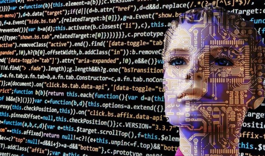
Three-dimensional imaging and contrast-enhanced mammography procedures are being performed with the mammography device located at the Senology Center of Izmir University of Economics (IUE) Medical Point Hospital. The images obtained are scanned by artificial intelligence, which acts as a second pair of eyes for the radiologist, leading to fast and reliable results in breast cancer diagnosis.
Early diagnosis saves lives in breast cancer. Technology and software are coming to the forefront for early diagnosis. The Senology Center at Izmir University of Economics (IUE) Medical Point Hospital aims to achieve fast and reliable results by using technology and artificial intelligence. While three-dimensional imaging and contrast-enhanced mammography procedures are applied with the mammography device used at the center, stereotactic biopsy and vacuum-assisted biopsy are performed for lesions seen as suspicious in both non-contrast and contrast-enhanced mammograms.

The images obtained with the device undergo a scan by software that has 92-94 percent sensitivity. Thanks to the software using artificial intelligence technology, the available images are compared with millions of benign and malignant lesion images. Artificial intelligence supports the radiologist as a second pair of eyes, helping to obtain fast and reliable results in breast cancer diagnosis.
Radiology Specialist Dr. Faculty Member Erkan Şahin said, “Our senology center uses a mammography device capable of taking three-dimensional tomosynthesis images. Three-dimensional imaging provides us with more information, especially in patients with dense breast tissue, and takes tomographic images with low-dose radiation. The device can also perform contrast-enhanced mammography procedures. Contrast-enhanced mammography is a serious alternative, especially for patients who cannot enter an MRI device for a breast MRI. With this device, in which we are pioneers in Izmir, we can also perform biopsies on lesions we see as suspicious in both non-contrast and contrast-enhanced mammograms without harming the patient, using both stereotactic and vacuum biopsy methods.”
Emphasizing that one of the biggest problems in breast imaging is pain during mammography, Dr. Faculty Member Erkan Şahin said, “The pain problem occurs due to compression during mammography. With this device, we have a mechanism that can give the compression control to the patient themselves. The patient compresses the breast themselves, and it hurts less.”
Stating that they use licensed FDA and CE-approved software after routinely obtaining the images, Şahin says the following:
“This software has 92-94 percent sensitivity. The artificial intelligence system recognizes every breast and provides a second perspective to the radiologist. Artificial intelligence stores millions of images of benign and malignant lesions proven by tissue diagnosis in its memory. It compares them with the mammography result in front of it and provides the result in a very short time. Artificial intelligence software is extremely important, especially for finding lesions hidden inside breast tissue, and it has a sensitivity of over 90 percent.”
News Source: İHA